Adhesion
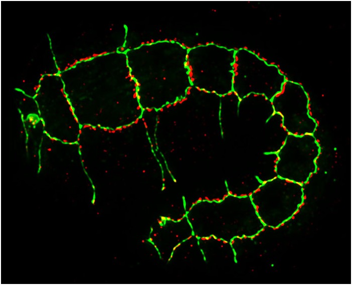
An elongating C. elegans embryo stained for HMP-1/α-catenin (green) and AJM-1 (red) [Jeff SImske].
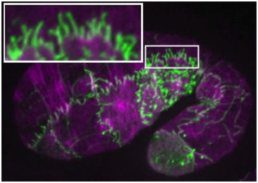
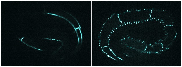
Morphogenesis requires that cells make strong connections with one another. We are studying how the cadherin complex, which is conserved in all metazoans, regulates morphogenesis. We are using a variety of approaches to study the cadherin complex, including:
(1) Structure-function in the embryo to identify important regions in cadherin complex proteins;
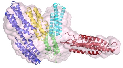
Our structure-function work has been based on actual structures of C. elegans proteins; much of our earlier work was done in collaboration with Hee-Jung Choi (Seoul National University) and BIll Weis’s laboratory (Stanford University).
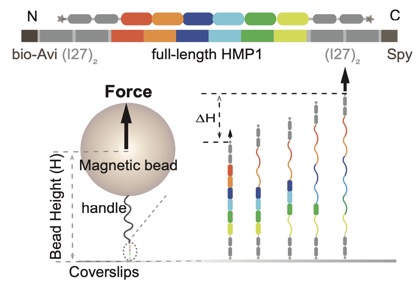
(2) Single-molecule force spectroscopy to study the mechanical properties of cadherin complex proteins in collaboration with Yan Jie’s group (National University of Singapore/Mechanobioloogy Institute) to identify functionally important domains required for mechanotransduction. For an early preview of this work see this bioXriv preprint: Link.
(3) Functional genomics and proteomics to identify proteins and pathways that act in concert with the cadherin complex during morphogenesis. These include actin-binding and junctional proteins and the Slit/Robo GAP, SRGP-1.